
RECOMMENDED APPLICATION
TEN is an ultra-premium nutritional supplement that supports your body’s mitochondria, which are the cell’s power producers.
TEN pills, taken with a full meal, are to be taken orally.
Adults should take one to two capsules per day.
Children under the age of 18 are not advised to use this product.
Consult your healthcare provider before using this or any other dietary supplement if you are pregnant or nursing, or if you have a medical condition.
If an allergic response occurs, stop taking it.
SUMMARY
The purpose of this study was to conduct pilot clinical testing on the effects of a new nutraceutical formula from Bod• Pro, Inc. on human mitochondria at the cellular level in order to help develop a clinical proof-of-concept trial.
A number of components in the nutraceutical formula Bode TEN are recognized to support mitochondrial activity.
Mitochondria are intracellular organelles that produce energy for the cell.
They are made up of membranous bodies that might take the form of oval bodies or a network of tubes.
As a result, rather of counting the number of mitochondria in a cell, the amount of mitochondria in a cell is measured as mitochondrial mass per cell.
PRODUCT DESCRIPTION
Ingredients & Supplement Facts
| Serving size: 1 capsule Serving per container: 30 | ||||
|---|---|---|---|---|
| Amount Per Serving | % Daily Value | |||
| Riboflavin | 12.5 | mg | 962 | % |
| Niacin (as niacinamide) | 50 | mg | 313 | % |
| Iron (as albion ferrochel) | 0.5 | mg | 3 | % |
| Magnesium (as magnesium citrate) | 25 | mg | 6 | % |
| Zinc (as zinc citrate) | 15 | mg | 136 | % |
| Selenium (as selenomethionine) | 200 | mcg | 364 | % |
| Copper (as copper citrate) | 2 | mg | 222 | % |
| Manganese (as manganese citrate complex) | 2 | mg | 87 | % |
| Molybdenum (as molybdenum citrate) | 100 | mcg | 222 | % |
| Proprietary Mitochondria Support Blend (CoEnzyme Q10 and PQQ Na2) | 60 | mg | * | |
| * Daily Value (%DV) Not Established |
Other Ingredients
TEN contains the following inactive carriers: Vegetable Capsule, Microcrystalline Cellulose, Silicon Dioxide, and Magnesium Stearate.
PRODUCT DESCRIPTION
TEN is a patent pending propriety formula that enhances cellular energy production, increases stamina and promotes antioxidant cellular protection from oxidative stress.*
MECHANISM OF ACTION AND BENEFITS
A number of proprietary components in the Bode TEN formula have been shown to support mitochondrial activity.
Mitochondria are intracellular organelles that produce 90 percent of the cellular energy in the body.
Bod• Pro chose to evaluate the impact of its patent-pending TEN formula on human mitochondria at the cellular level and general well-being in a pilot clinical trial.
Six pilot tests have been carried out to date in order to assess the effects of Bode TEN at the cellular level, particularly in stressful human culture conditions.
IN VITRO TESTING OF PRODUCTION OF MITOCHONDRIA AND ANTIOXIDANT CAPACITY. The TEN formula significantly increased mitochondrial mass after 2 hours in cells following treatment with the product and showed robust antioxidant capacity.
HUMAN CLINICAL STUDY ON PRODUCTION OF MITOCHONDRIA AND INCREASING HEALTHY MITOCHONDRIA FUNCTION. In a small-scale independent, randomized, double blind placebo-controlled clinical study, twelve test subjects were randomly divided into two groups and received TEN or a placebo for 30 days. After the 30-day evaluation period, the study showed that those who consumed TEN: (i) had a rapid 20% increase in mitochondria over baseline and 32% increase after 4 weeks; and (ii) a significant increase of 14% in the functional capacity/energetics of the mitochondria within 2 hours of consumption.*
IN-VITRO STUDY ON PROTECTION OF MITOCHONDRIAL FUNCTION FROM OXIDATIVE STRESS AND INFLAMMATION SUMMARY. The TEN formula supported mitochondrial metabolic activity under normal, oxidative stress and inflamed culture conditions.*
IN VITRO TESTING OF MITOCHONDRIAL MARKERS AND INHIBITION OF FREE RADICAL STRESS. The TEN formula protected cells from oxidation under oxidative stressed culture conditions supported mitochondrial membrane potential under normal oxidative stress and inflamed culture conditions and demonstrated anti-inflammatory effect.*
HUMAN STUDY ON THE EFFECTS OF TEN ON CORTISOL LEVELS. In a twelve-week human study, ten patients were given a double dose of TEN for a period of 12 weeks. Blood draws were taken at week 0 and week 12. The results indicated that TEN may support healthy cortisol levels already within the normal range.*
HUMAN STUDY ON THE EFFECTS OF TEN ON DHEA LEVELS. Ten patients who were given a double dose of TEN for a period of 12 weeks were also tested for their DHEA levels. Blood draws were taken at week 0 and week 12. The results indicated that TEN may support healthy DHEA levels already within the normal range.*
Bode Pro TEN: https://www.pdr.net/full-prescribing-information/Ten-dietary-supplement-24294
Despite the fact that these six investigations were small in scope and yielded no definitive outcomes, their findings are incredibly promising.
The data from these exploratory tests will be used by Bod• Pro to undertake a bigger human clinical research in the future.
CLINICAL EXPERIENCE
1. In Vitro Production of Mitochondria and Increasing Healthy Mitochondria Function
EXECUTIVE SUMMARY
After 2 hours, cells treated with an aqueous portion of the TEN nutraceutical formula had dramatically increased mitochondrial mass.
The timeframe (2 hours) is favorable for future human clinical research that will include the mitochondrial mass assay.
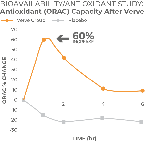
Pilot research was also conducted on TEN’s total antioxidant capacity (TAC) in order to determine the antioxidant levels present in an aqueous version of the TEN formula.
This is pertinent to the antioxidants that are likely to be accessible in future clinical investigations after human ingestion.
RESULTS
2 MAJOR STUDY ENDPOINTS
The capacity of a chemical to generate an increase in the number of Mitochondrial organelles within human leukocytes is known as Mitochondrial Mass Per Cell (MMPC) (white blood cells).
Total Antioxidant Capacity (TAC) is defined as a compound’s capacity to protect human leukocytes from free radical damage.
ENDPOINTS
- Mitochondrial Mass Per Cell:
- Increases in MMPC were detected at 2 hours following treatment of cells with the TEN formula (see graphs below).
- The 0.125 g/L dose of the TEN formula led to an 80-140% increase in MMPC in 3 different white blood cell populations. This shows the relative potency of the TEN formula, which is promising for future clinical studies where mitochondrial mass per cell will be tested in blood samples from human subjects before and after consuming the TEN formula.


Total Antioxidant Capacity:
- The TEN formula showed robust antioxidant capacity. This is important since it is expected that many of the water-soluble compounds present in the TEN formula will be readily available following human consumption.
- Graph below shown in Gallic acid equivalents.

2. Human Clinical Study of Mitochondria and Increasing Healthy Mitochondria Function
A placebo-controlled cross-over study design was used for the testing of acute effects, following the design of previous human testing of cellular effects of natural products.
This project involved 12 people, where each person’s acute response was tested on the active product compared to a placebo. This was followed by a 4-week open-label phase.
The volunteers were tested on separate clinic days with 7 days’ wash-out period between test days. The sequence for testing of acute effects of the TEN formula and placebo, as presented below, is an example only, since the order in which products were fed to study participants for acute effects was randomized:
SUBJECTS
12 volunteers were recruited for this study.
INCLUSION CRITERIA
- Healthy adult volunteers of either gender.
- Age 45-70 years.
- BMI between 25 – 40.
- Veins easy to see in both arms.
PRESCREENING
The pre-screening involved an interview to document gender, age, BMI, medical/surgical history, diet/lifestyle, current health issues, medication, and supplement use.
STUDY VISITS
On each test day, our previously established protocol for acute effects of the TEN formula was applied. This involved careful management of the human subjects as well as the study environment, and a full hour of quiet resting before the baseline blood draw:
Upon arrival on the morning of each clinic day, participants rested in a seated position quietly for 1 hour prior to baseline blood draw. This resting period is crucial to gain representative baseline data. During this time, questionnaires were completed to monitor previous meals, snacks, exercise, stressors, and recent sickness. The baseline blood draw was followed immediately by feeding a product to the volunteer, who continued to rest quietly. One more blood draw was taken at 2 hours after consumption of the TEN formula. Blood pressure and responses to a brief questionnaire on mental state were recorded prior to each blood draw.
Blood samples were tested on clinic days for mitochondrial mass and activity. Blood samples were also sent for testing for Complete Metabolic Panel (CMP) and Complete Blood Count (CBC) at the baseline and 4 week visits, and high-sensitivity C- reactive protein (hsCRP) testing was done on both blood draws from each acute clinic day as well as the 4 week visit.
Serum was collected on all blood draws both for testing of a panel of inflammatory cytokines (this report) and for banking for future testing.
Questionnaires were administered at each visit. This included a questionnaire on general health and wellbeing (29 questions) and a questionnaire pertaining to fatigue (14 questions).
DEMOGRAPHICS
OF THE STUDY POPULATION
Table 1. Demographics of study participants.
| Females: | 7 | Males: | 5 |
| Age average* | 59.1 ± 5.0 | Age average* | 57.7 ± 9.6 |
| Age range | 53.7 – 66.9 | Age range | 47.1 – 65.8 |
| BMI average | 27.5 ± 4.9 | BMI average | 30.2 ± 2.3 |
| BMI range | 22.9 – 33.0 | BMI range | 27.3 – 33.0 |
COMPLIANCE
Compliance was evaluated by capsule count for product returned at the end of the 4-week open-label part of the study. The average compliance during the study was 96% (range 84- 100%).
SOURCE OF TEST PRODUCTS
The active product was provided by the study sponsor. The TEN formula is a blend of vitamins and nutritional ingredients designed to enhance mitochondrial mass and function. Study participants were given 4 weeks supply of the test product. This product was also used for the placebo-controlled cross-over part of the study.
Placebo (used for the acute phase of the study) consisted of rice flour encapsulated in veggie capsules.
BLINDING OF PRODUCT AND PLACEBO FOR TESTING OF ACUTE EFFECTS
Volunteers were randomized so that on each of the clinic visits where acute effects were measured, half of the volunteers received placebo and the other half received active product. On their subsequent clinic visit to measure acute effects, volunteers received the remaining product (placebo or product), which they had not previously received.
Prior to the acute study days, TEN product and placebo were blinded and randomly assigned to each volunteer’s first or second acute study day. Two people, neither of whom was directly involved in administering product to study participants, witnessed this. Clinic personnel and study participants were not aware of the identity of the product being administered on a particular study day.
METHODS AND DATA ANALYSIS
The testing described herein was performed on white blood cells (leukocytes) isolated from peripheral blood drawn from study participants taken before consumption, 2 hours post consumption and after 4 weeks of consumption. 3 different cell lines were used in the study.
Blood cell populations:
- Lymphocytes are a type of white blood cell that are round in shape with a round nucleus that occupies the majority of the cell. Three types of cells belong to the lymphocyte class. These are natural killer cells, T lymphocytes and B lymphocytes.
- Monocytes are the largest type of white blood cell. They are present in the blood circulation, and after leaving the blood and migrating into tissue they become macrophages and serve a crucial link between the innate and adaptive immune responses.
- Neutrophils are also known as polymorphonuclear (PMN) cells because they contain a nucleus whose shape (morph) is irregular and contains many (poly) lobes. They are the most abundant type of white blood cell and are the first line of defense for bacterial infection. Neutrophils engulf and kill bacteria by releasing enzymes stored in intracellular granules. Neutrophils are also involved in inflammation, including migrating to sites of tissue injury in response to chemical signals.
Group averages and standard error of the means (SEM) for each data set were calculated using Microsoft Excel. Statistical significance of changes from baseline to later assessments was evaluated by between-treatment analysis using ‘within-subject’ analysis using the two-tailed, paired t-test. The probability (p) values indicate the strength of the evidence that the study-specific ‘treatment’, i.e. the TEN formula consumption, had an effect that can be considered statistically significant. On the tables and data graphs, the levels of statistical significance are displayed with asterisks following the format below. Levels of significance:
- When a change in the study reached a high level of statistical significance, as defined as p<0.01 (1% risk of error; 99% confidence interval), this is indicated with “**”.
- When a change in the study reached statistical significance as defined as p<0.05 (5% risk of error; 95% confidence interval), this is indicated with “*”.
- When a change in the study revealed only a marginal effect, this is indicated as a statistical trend. A trend is defined as p<0.10 (10% risk of error; 90% confidence interval), and indicated with “(*)”.
RESULTS
STUDY ENDPOINTS
- Mitochondrial Biogenesis/Mass: defined as, the ability of a compound to induce natural synthesis or an increase in volume of Mitochondrial organelles in the human body.
- Mitochondrial Energetics/Function: defined as, the ability of a compound to induce ATP or energy synthesis in Mitochondrial organelles in the human body.
- Study Participant Energy Questionnaire: defined as, a standardized testing method used to evaluate participant’s physical energy, mental focus and mental function after ingesting a compound versus placebo.
- Safety Parameters and Inflammatory Markers: defined as, a Complete Blood Count (CBC), Complete Metabolic Profile (CMP) and endogenous inflammatory cytokines. Used to evaluate the relative safety of a compound when ingested by humans over short and long term use.
4 MAJOR STUDY ENDPOINTS
1. MITOCHONDRIAL BIOGENESIS/MASS:
- Acute/Short-Term Results (2 hours):
- Summary: Over the two hours of inactive rest on the acute study days, increases in mitochondrial mass per cell were seen in all blood cell populations following consumption of the TEN formula. The increase associated with the TEN formula consumption reached a statistical trend (p<0.10) in the lymphocyte population, when compared to baseline levels.
- A rapid 20% increase (average) in the volume of mitochondria from baseline and a statistically significant 5% increase greater than placebo.
- Long-Term Results (4 weeks):


2. MITOCHONDRIAL ENERGETICS/FUNCTION:
- Acute/Short-Term Results:
- Summary: In the case of the PMN cell population, an increase in mitochondria energetics was seen post TEN formula consumption. This suggests that the consumption of the TEN formula triggered an increase in metabolic activity in contrast to a reduction seen in activity following placebo consumption.
- A 14% increase in the functional capacity/energetics of the mitochondria was seen with 2 hours of consumption (shown in table below). There was no difference between placebo and active test product. The reduction may relate to people sitting still for two hours.
Table 3. Acute effects on mitochondrial function in PMN cells (MTT assay)
| Placebo | Active Test Product | |||||||
|---|---|---|---|---|---|---|---|---|
| Baseline* | 2 hours* | % change | P-value | Baseline* | 2 hours* | % change | P-value | P-value (placebo/MEF-2) |
| 0.83 ± 0.10 | 0.72 ± 0.08 | -14.05% | 0.2757 | 0.67 ± 0.07 | 0.76 ± 0.07 | 13.62% | 0.2757 | 0.1234 |
| * average ± SEM |
3. STUDY PARTICIPANT ENERGY QUESTIONNAIRE
- Energy questionnaire evaluated effects on current mental state and used a visual analog scale (VAS) with a sliding scale from 0-100. This questionnaire was administered prior to all blood draws.
- Acute/Short-Term Results:
- Summary: An improvement in physical energy levels and mental focus and mental functioning was seen 2 hours after consuming the TEN formula. A similar increase was not seen after consuming placebo, where a slight decrease was seen for these scores. When comparing an individuals’ response to the TEN formula versus placebo, statistical significance (p<0.05) was obtained in mental focus and mental functioning following the TEN formula consumption (see table below).
| Placebo | MEF-2 | Placebo/MEF-2 Comparison * | |||||
|---|---|---|---|---|---|---|---|
| Questions | Baseline† | 2 hours† | P-value‡ | Baseline† | 2 hours† | P-value‡ | P-value‡ |
| What is your current energy level? | 68.25 ± 7.96 | 65.00 ± 8.34 | 0.7460 | 65.75 ± 8.38 | 74.42 ± 8.03 | 0.0479* | 0.1242 |
| How focused do you feel? | 82.17 ± 6.06 | 72.00 ± 8.53 | 0.2064 | 81.33 ± 5.56 | 85.00 ± 5.14 | 0.4870 | 0.0213* |
| What is your current level of mental functioning? | 80.00 ± 6.28 | 71.08 ± 7.61 | 0.2097 | 79.42 ± 6.08 | 82.58 ± 5.92 | 0.5926 | 0.0247* |
| What is your current stress level? | 8.08 ± 2.61 | 1.67 ± 0.74 | 0.0253* | 1.50 ± 0.62 | 1.58 ± 0.56 | 0.7774 | 0.8864 |
| What is your current level of relaxation? | 81.42 ± 4.98 | 78.25 ± 10.61 | 0.7739 | 81.58 ± 8.20 | 86.50 ± 7.90 | 0.6682 | 0.3514 |
| * Comparison between placebo and MEF-2 at 2 hours † average ± SEM ‡ p<0.05 is indicated with “*”. |
Long Term Results
- A slight improvement in energy levels and mental focus/functioning was seen following 4 weeks’ consumption of the TEN formula (see table below).
Table 14. Energy Questionnaire — Long-term effects
| Questions | Baseline 1* | Baseline 2* | P-value | 4 weeks* | P-value†, ‡ | P-value§ |
|---|---|---|---|---|---|---|
| What is your current energy level? | 61.50 ± 8.53 | 72.00 ± 7.45 | 0.2525 | 77.08 ± 6.67 | 0.0824* | 0.0437 |
| How focused do you feel? | 81.33 ± 6.27 | 82.00 ± 5.32 | 0.8628 | 82.25 ± 5.99 | 0.8262 | 0.9750 |
| What is your current level of mental functioning? | 78.58 ± 6.49 | 80.83 ± 5.83 | 0.6270 | 82.17 ± 5.20 | 0.3881 | 0.7106 |
| What is your current stress level? | 4.92 ± 2.12 | 4.67 ± 2.16 | 0.9331 | 16.50 ± 6.66 | 0.1528 | 0.1171 |
| What is your current level of relaxation? | 83.33 ± 4.64 | 79.67 ± 8.37 | 0.6684 | 81.83 ± 4.91 | 0.8162 | 0.8164 |
| * average ± SEM † p<0.01 is indicated with “*”. ‡ Comparison from Baseline 1 to 4 weeks. § Comparison from Baseline 2 to 4 weeks. |
4. SAFETY PARAMETERS AND INFLAMMATORY MARKERS:
- Blood chemistry and complete blood count:
- Values were within normal ranges at study start and following 4 weeks’ consumption of the TEN formula. Slight but statistically significant increases were seen for alkaline phosphatase, albumin, globulin and total protein.
- Safety testing – Inflammatory markers short-term effects:
- Hs-CRP levels in serum remained constant following both placebo and MEF-2 consumption.
- The cytokines IL-2, IL-4, IL-6 and IL-8 were undetectable in baseline serum samples and remained so two hours following placebo or the TEN formula consumption.
- No significant changes to levels of IL1β (detectable at baseline in only one volunteer) IFN-γ or TNF-α were seen following either placebo or the TEN formula consumption.
- A significant decrease in MCP-1 serum levels in all volunteers’ serum was seen following placebo consumption (p<0.01) but not following the TEN formula consumption.
- Safety testing – Inflammatory markers long-term effects:
- Hs-CRP levels in serum showed nonsignificant decreases in one half of the volunteers and non-significant increases in the other half following 4 weeks’ consumption of the TEN formula.
- The cytokines IL-2, IL-4, IL-6 and IL-8 were undetectable in baseline serum samples and remained so following 4 weeks’ consumption of the TEN formula.
- IFN-γ levels in serum remained constant while IL-1β (detectable at baseline in only one volunteer), TNF-α and MCP-1 levels slightly decreased following 4 weeks’ the TEN formula consumption.
Disclosures
These statements have not been evaluated by the Food and Drug Administration. This product is not intended to diagnose, treat, cure or prevent any disease.
3. Protection of Mitochondrial Function from Oxidative Stress and Inflammation Summary
EXECUTIVE SUMMARY
Bod•ē Pro distributes ultra-premium nutritional supplements for wellness and cellular nutrition. Bod•ē Pro’s latest offering, TEN, which is focused on supporting cellular energy through support of mitochondrial function. This pilot project has generated results to support the effects of TEN at the cellular level, particularly under stressed culture conditions.
TEN provided cellular antioxidant protection, showing that it contains antioxidants capable of penetrating into cells and protecting them from the inside-out, when the cells were exposed to free radical stress.*
When cells are stressed by free radicals and by inflammatory conditions, the mitochondria suffer. Testing applied several types of stressors to cells to test the protection offered by TEN. TEN showed multiple levels of support of mitochondrial in bioassays:
- TEN supported mitochondrial metabolic activity under normal, oxidative stress, and inflamed culture conditions.*
- TEN supported healthy mitochondrial mass per cell under inflamed conditions in the presence of the TEN formula.*
The data are also immediately useful for planning of the fourth clinical trial protocol on Bod•ē Pro TEN. The data reported here points to inflammation as the most useful stressor for testing protective effect of TEN.
PURPOSE
The purpose of this pilot project was to test the protective effects of TEN on mitochondrial function under various experimental conditions of oxidative stress, inflammation, and reduced oxygen delivery, using cellular bioassays.
The results from the comparison of various culture conditions, to cause stress on mitochondrial function, will help plan which test conditions to use in the subsequent clinical trial, which needs to be initiated as soon as possible.
BACKGROUND
A novel nutraceutical formula, TEN has been developed for the support of mitochondrial function, with ingredients backed by research.
A key ingredient is pyrroloquinoline quinone (PQQ), which has shown rapid effects on inflammation and mitochondrial-related metabolism in human subjects, with peak serum levels at 2 hours.i Therefore, the planned clinical study aims at documenting effects within 1-2 hours after consuming a single dose of product, compared to placebo.
Another key ingredient is Coenzyme Q10, also well-known to support mitochondrial function under stressful conditions.ii iii
The effect of the patent pending blend of mitochondria-targeted ingredients needs to be documented, both under normal conditions, and under conditions of specific types of stress, reflecting situations simulating exercise-induced stress, as well as inflammation.
METHODS
TESTS PERFORMED
This pilot project compared several tests that evolve around:
- Total antioxidant capacity.
- Cellular antioxidant protection due to uptake of antioxidant compounds.
- Mitochondrial volume per cell:
- Under normal culture conditions (unstressed).
- Under oxidative stress.
- Under inflamed conditions.
- Mitochondrial metabolic activity:
- Under normal culture conditions (unstressed).
- Under oxidative stress.
- Under inflamed conditions.
PRODUCT HANDLING
The formula TEN contains a blend of water-soluble and water-insoluble/poorly soluble compounds. Therefore, this pilot testing compared the cellular effects of three different methods of handling the product for introduction into the liquid nutrient-rich medium in which the cells are grown in cell culture.
| Extraction/handling | Solvent for making stock | Serial dilutions | Proportion |
|---|---|---|---|
| TEN Aqueous handling | Physiological saline | Physiological saline | |
| TEN Ethanol handling | Ethanol 95% (blend) | Physiological saline | |
| TEN Aqueous/ethanol blend | (blend) | Physiological saline | 50:50 blend |
RESULTS
TOTAL ANTIOXIDANT CAPACITY
The three extracts shown in Table 1 were tested in the Folin-Ciocalteu assay (also known as the total phenolics assay). This assay makes use of the Folin-Ciocalteu reagent to measure antioxidants. The assay is performed by adding the FolinCiocalteu’s phenol reagent to serial dilutions of each extract, thoroughly mixing, and incubating for 5 minutes. Sodium carbonate is then added, starting a chemical reaction producing a color. The reaction is allowed to continue for 30 minutes at 37°C. Optical absorbance is measured at 765nm in a colorimetric plate reader. Gallic acid is used as a reference standard, and the data reported in Gallic Acid Equivalents per gram product. Data from this assay helps interpret the subsequent Cellular Antioxidant Protection assay, as well as the assays for mitochondrial function. The results confirmed that TEN contains antioxidants. The antioxidant capacity was greater in the water-soluble fraction than in the ethanol fraction.
CELL-BASED ANTIOXIDANT PROTECTION (CAP-E) ASSAY
The rationaleiv behind the method that we use is important: It allows assessment of anti- oxidant potential in a method that is comparable to the ORAC antioxidant test, but only allows measurement of antioxidants that are able to cross the lipid bilayer cell membrane, enter the cells, and provide biologically meaningful antioxidant protection under conditions of oxidative stress. We developed the CAP-e bioassay specifically to work with natural products and ingredients.v The method has been used on multiple types of natural products and ingredients, published in the peer-reviewed scientific literature.vi vii viii ix x xi
As a model cell type, we use the red blood cell (RBC). This is an inert cell type, in contrast to other cell types. We developed this assay particularly to be able to assess antioxidants from complex natural products in a cell-based system.
Freshly purified human RBC are washed repeatedly in physiological saline, and then exposed to the test products. During the incubation with a test product, any antioxidant compounds able to cross the cell membrane can enter the interior of the RBC. Then the RBC are washed to remove compounds that were not absorbed by the cells, and loaded with the DCF-DA dye, which turns fluorescent upon exposure to reactive oxygen species. Oxidation is triggered by addition of the peroxyl free radical generator AAPH. The fluorescence intensity is evaluated. The low fluorescence intensity of untreated control cells serve as a baseline, and RBC treated with AAPH alone serve as a positive control for maximum oxidative damage.
When we observe a reduced fluorescence intensity of RBC exposed to a test product and subsequently exposed to AAPH, this indicates that the test product contains antioxidants available to penetrate into the cells and protect these from oxidative damage.
The results showed that water-soluble antioxidants in TEN were capable of entering into and protecting the cells from oxidative stress-induced intracellular damage.
MITOCHONDRIAL METABOLIC ACTIVITY WHEN EXPOSED TO VARIOUS STRESSORS
The testing described below was performed on freshly isolated peripheral blood mononuclear cells from a healthy human blood donor. Serial dilutions of the test product (using all 3 handling methods in parallel) were added to cell cultures, where the cultures were tested for mitochondrial metabolic activity, using the MTT assay. Freshly purified cells were cultured for 24 hours in the presence of TEN, after which MTT reagents were added to allow a color formation to take place in proportion to mitochondrial function. In the MTT bioassay, chemical reactions trigger a specific color development based on cellular functions:
- When a reduction in color is measured, this is linked to a reduced cellular viability, either as a result of direct killing, or inhibition of mitochondrial function.
- When an increase in color is measured, this has multiple possible explanations: 1) increased cell numbers (growth); 2) increased mitochondrial mass; and 3) increased mitochondrial function (energy production).
Under the test conditions used for this project, no cellular growth was expected, therefore the changes are proportional to the combination of increased mitochondrial mass, and increased mitochondrial metabolic activity.
Culture conditions
One set of cell cultures was allowed to incubate under normal cell culture conditions (“Normal culture”), without any added stressors. The following four sets of cell cultures were made in parallel:
- Normal culture.
- Oxidative stress (induced by adding H2O2).
- Inflammation (induced by LPS (a highly inflammatory bacterial endotoxin)).

MITOCHONDRIAL MASS PER CELL
The testing described below was performed on freshly purified white blood cells from a healthy human blood donor. Serial dilutions of the test product (using all 3 handling methods in parallel) were added to cell cultures, where the cultures were tested for mitochondrial volume after 2 hours incubation (to mimic timing proposed for clinical study). Untreated control samples were processed in parallel.
Cell types
Different cell types have different mitochondrial activity levels, both under normal and under stressed conditions. During flow cytometric analysis, separate analysis was performed on the following three cell types in the blood sample:
- Lymphocytes (Ly): Fairly inactive in the types of cell cultures used here;
- Monocytes (Mo): Moderately active cells, highly reactive to stressors;
- Polymorphonuclear cells (PMN): Highly active cells, extremely reactive to stressors.

Culture conditions
One set of cell cultures was allowed to incubate under normal cell culture conditions (“Normal culture”), without any added stressors. The following four sets of cell cultures were made in parallel:
Normal culture: Moderate increases in mitochondrial mass per cell when cells were treated with the aqueous as well as the ethanol-extract of TEN.
Oxidative stress (induced by adding H2O2): Mild increases in mitochondrial mass per cell when Mo and PMN cells were treated with the ethanol-extract of TEN.
Inflammation (induced by LPS (a highly inflammatory bacterial endotoxin)): TEN offered strong protection of mitochondrial mass per cell for all three cell types.
After incubation, cells were stained with a fluorescent probe that stains mitochondria in proportion to mitochondrial volume per cell. The overall sum of fluorescent output per cell is a measure of mitochondrial volume per cell. The Attune® acoustic aligning flow cytometer tracks cell numbers per volume unit, and provides fluorescence intensity measurements per cell.
On the following pages are two sets of graphs for each culture condition and cell type. The first graph shows the TEN-induced change for all three test fractions for all three doses tested.
The second graph serves to illustrate the effects of TEN when compared to untreated versus stressed cells.


This pilot project has generated results to support the effects of TEN at the cellular level, particularly under stress human culture conditions.
TEN supported mitochondrial metabolic activity under normal, oxidative stress and inflamed human culture conditions.
TEN supported healthy mitochondrial mass per cell under specific culture conditions: Mitochondrial mass per cell was mildly supported by TEN under normal conditions and strongly supported under inflamed conditions. The human cultures under oxidative stress showed some support by TEN for the more active cells (monocytes and PMN cells).
Disclosures
These statements have not been evaluated by the Food and Drug Administration. This product is not intended to diagnose, treat, cure or prevent any disease.
4. IN VITRO TESTING OF MITOCHONDRIAL MARKERS AND INHIBITION OF FREE RADICAL STRESS
1. PURPOSE
The purpose of this project was to test the effects of TEN on cellular reduced glutathione levels and mitochondrial membrane potential under both normal and oxidative stress culture conditions, and to test TEN’s effect on inhibiting free radical stress in human inflammatory cells.
This project is a direct continuation of previous testing.
The effects of the blend of mitochondria-targeted ingredients were documented, both under normal conditions, and under conditions of specific types of stress, reflecting situations simulating exercise-induced stress, as well as inflammation.
2. WORK PERFORMED
2.1 TESTS PERFORMED
This pilot project applied multiple tests to the increased understanding of TEN:
- Effect of product on intracellular levels of reduced glutathione under oxidative stress;
- Effect of product on mitochondrial membrane potential under normal and stressed culture conditions;
- Effect of product on free radical formation by human inflammatory cells under conditions of oxidative stress.
2.2 PRODUCT HANDLING
The energy formula TEN contains a blend of water-soluble and water-insoluble/poorly soluble compounds. Therefore, this pilot testing compared the cellular effects of three different methods of handling the product for introduction into cell culture.
| Extraction/handling | Solvent for making stock | Serial dilutions | Proportion |
|---|---|---|---|
| TEN Aqueous handling | Physiological saline | Physiological saline | |
| TEN Ethanol handling | Ethanol 95% (blend) | Physiological saline | |
| TEN Aqueous/ethanol blend | (blend) | Physiological saline | 50:50 blend |
| Ethanol control | Ethanol 95% | Physiological saline |
3. DATA PRESENTATION
Data is presented in the Results section for each of the 4 bioassays following this system:
- An initial bar graph shows the most important data set for a particular bioassay, displayed as the percent change from control cultures;
- This is followed by a set of line graphs where all 4 doses of all 4 products are compared on single graphs, displayed as percent change from control data;
- Raw data is shown as bar graphs with any statistical significance indicated with asterisks. Each product is plotted separately with its appropriate control;
- For the reduced glutathione and mitochondrial membrane potential assays, several cell types were analyzed, and data is presented sequentially for individual cell populations (lymphocytes and monocytes for reduced glutathione; lymphocytes, monocytes and PMN cells for mitochondrial membrane potential).
4. RESULTS
4.1 INTRACELLULAR GLUTATHIONE LEVELS IN HUMAN CELLS
Glutathione (GSH) is an important antioxidant in plants, animals, fungi, and some bacteria. Glutathione is capable of preventing damage to important cellular components caused by damaging free radicals. Glutathione exists in two states: reduced (GSH) and oxidized (as the molecule glutathione disulfide, GSSG). In the reduced state, the thiol group of cysteine is able to donate a reducing equivalent to other molecules, such as reactive oxygen species, to neutralize the free radicals. We used an established protocoliv for flow cytometric evaluation of cellular protection by TEN, where product was applied to human peripheral blood mononuclear cells in the absence versus presence of free radical stress. ThiolTracker™ Violet is a bright intracellular thiol probe for reproducible detection of intracellular levels of reduced glutathione.v Since reduced glutathione represents the majority of intracellular free thiols in the cell; ThiolTracker™ Violet can be used to estimate the cellular level of reduced glutathione by flow cytometry.
Human peripheral blood mononuclear cells were treated with test products for 30 minutes, after which test products were removed. One set of cell cultures was handled using normal culture conditions (no stressors) while the other set of cell cultures was exposed to 1 mM H2O2 for 1 hour (oxidative stress culture conditions). Cells were then stained with ThiolTracker™ Violet and acquired by flow cytometry using an Attune® acoustic focusing cytometer. Data were analyzed for changes in fluorescence intensity, which reflects the reduced glutathione levels in the cells.
Cell types
During flow cytometric analysis, separate analysis was performed on the following two cell types in the blood sample:
- Lymphocytes (Ly): Fairly inactive in the types of cell cultures used here; Monocytes (Mono): Moderately active cells, highly reactive to stressors.

Figure 1. Flow cytometry analysis of cellular reduced glutathione levels. Electronic gates are set to define lymphocytes (Ly) and monocytes (Mono) based on their forward scatter (size) and side scatter (granularity) properties. The fluorescence intensity of the ThiolTracker™ Violet dye was measured for each cell type.
SYNOPSIS
Note: When reviewing this set of data, an increase in fluorescence intensity is a positive finding, and shows protection of reduced glutathione.
- Normal culture conditions
- Treatment of cells with products led to some slight reduction in cellular reduced glutathione levels;
- Monocytes were more sensitive to this effect, particularly at the 2 g/L dose of TEN Aqueous.
- Oxidative stress culture conditions
- Pretreatment of cultures with TEN Aqueous and TEN Ethanol prior to exposure to 1 mM H2O2 led to protection from loss of reduced glutathione*;
- For lymphocytes, protection was seen with the 3 highest doses of TEN Aqueous and the 0.4 and 0.08 g/L doses of TEN Ethanol*;
- Monocytes showed a reduction in cellular reduced glutathione when exposed to the 2 g/L dose of TEN Aqueous but were protected from loss of reduced glutathione by the 0.4 g/L dose of both TEN Aqueous and TEN Ethanol.*
- Immediately below is shown graphs for the protective results for 0.4 g/L TEN, specifically for the oxidative stress conditions.
- On subsequent pages the complete dose curves are shown, followed by raw data graphs.
- Pretreatment of cultures with TEN Aqueous and TEN Ethanol prior to exposure to 1 mM H2O2 led to protection from loss of reduced glutathione*;


6.1.1 RAW DATA – NORMAL CULTURE CONDITIONS
6.1.1.1 LYMPHOCYTES
- An initial bar graph shows the most important data set for a particular bioassay, displayed as the percent change from control cultures;
- This is followed by a set of line graphs where all 4 doses of all 4 products are compared on single graphs, displayed as percent change from control data;
- Raw data is shown as bar graphs with any statistical significance indicated with asterisks. Each product is plotted separately with its appropriate control;
- For the reduced glutathione and mitochondrial membrane potential assays, several cell types were analyzed, and data is presented sequentially for individual cell populations (lymphocytes and monocytes for reduced glutathione; lymphocytes, monocytes and PMN cells for mitochondrial membrane potential).

6.1.1.2 MONOCYTES

6.1.2 RAW DATA – OXIDATIVE STRESS CULTURE CONDITIONS
6.1.2.1 LYMPHOCYTES

6.1.2.2 MONOCYTES

6.2 MITOCHONDRIAL METABOLIC MEMBRANE POTENTIAL WHEN EXPOSED TO VARIOUS STRESSORS
JC-1 is a cationic carbocyanine dye that accumulates in mitochondria. The dye exists as a monomer at low concentrations and yields green fluorescence, similar to fluorescein. At higher concentrations, the dye forms J-aggregates that exhibit a broad excitation spectrum and an emission maximum at ~590 nm. These characteristics make JC-1 a sensitive marker for mitochondrial membrane potential. Healthy mitochondria will fluoresce in the orange spectrum, whereas stressed mitochondrial will fluoresce in the green spectrum.
The testing was performed on freshly purified white blood cells from a healthy human blood donor. Serial dilutions of the test product (using all 3 handling methods in parallel) were added to cell cultures, where the cultures were tested for mitochondrial membrane potential after 2 hours incubation (to mimic timing proposed for clinical study). Untreated control samples were processed in parallel.
One set of cell cultures was handled using normal culture conditions (no stressors). Two additional sets of cell cultures were made in parallel, where:
Oxidative stress was induced by adding H2O2;
Inflammation was induced by adding LPS (a highly inflammatory bacterial endotoxin).
Flow cytometric analysis for the mean fluorescence intensity in both the green and red spectrum were evaluated and the ratio between red (healthy membrane potential) and green (compromised membrane potential) fluorescence was used as a measure of relative mitochondrial membrane potential.
Cell types
Different cell types have different mitochondrial activity levels, both under normal and under stressed conditions. During flow cytometric analysis, separate analysis was performed on the following three cell types in the blood sample:
Lymphocytes (Ly): Fairly inactive in the types of cell cultures used here;
Monocytes (Mo): Moderately active cells, highly reactive to stressors;
Polymorphonuclear cells (PMN): Highly active cells, extremely reactive to stressors.

SYNOPSIS
Note: When reviewing this set of data, an increase in fluorescence intensity is a positive finding, and shows protection of the mitochondrial membrane potential.
- Normal culture conditions
- Treatment of cells with products led to increases in mitochondrial membrane potential*;
- Treatment with TEN Aqueous led to consistent increases in both lymphocyte and monocyte mitochondrial membrane potential.*
- The effects of the TEN Ethanol is inconclusive because higher increases were seen in cultures exposed to ethanol alone (Ethanol control)*;
- For PMN cells, the highest increases in mitochondrial membrane potential were seen in cultures treated with TEN Ethanol and TEN Blend.* The increases were seen with all 4 doses and they were higher than those seen with the Ethanol control.
- Treatment of cells with products led to increases in mitochondrial membrane potential*;
- Oxidative stress culture conditions
- Treatment of lymphocytes with all 4 doses of TEN Aqueous, TEN Ethanol and TEN Blend led to significant increases in mitochondrial membrane potential*;
- Treatment of monocytes with TEN Blend and ethanol alone (Ethanol control) led to higher increases in mitochondrial membrane potential than treatment with either TEN Aqueous or TEN Ethanol*;
- Treatment of PMN cells with TEN Aqueous led to decreased mitochondrial membrane potential at the two highest doses while the two lowest doses and all 4 doses of TEN Blend led to increases in mitochondrial membrane potential.*
- Inflammatory culture conditions
- Treatment of lymphocytes with the two highest doses of TEN Aqueous led to significant increases in mitochondrial membrane potential*;
- Treatment of monocytes with products did not lead to increased mitochondrial membrane potential;
- Treatment of PMN cells with the two highest doses of TEN Aqueous and the highest dose of Ethanol control led to decreases in mitochondrial membrane potential while treatment with the two highest doses of TEN Blend and the three lowest doses of Ethanol control led to slight increases.*
Immediately below is shown graphs for the protective results for 0.4 g/L TEN, specifically for the oxidative stress conditions. On subsequent pages the complete dose curves are shown, followed by raw data graphs.


6.2.1 RAW DATA – NORMAL CULTURE CONDITIONS
6.2.1.1 LYMPHOCYTES

6.2.1.2 MONOCYTES

6.2.1.3 PMN CELLS

6.2.2 RAW DATA – OXIDATIVE STRESS CULTURE CONDITIONS
6.2.2.1 LYMPHOCYTES

6.2.2.2 MONOCYTES

6.2.2.3 PMN CELLS

6.2.3 RAW DATA – INFLAMMATORY CULTURE CONDITIONS
6.2.3.1 LYMPHOCYTES

6.2.3.2 MONOCYTES

6.2.3.3 PMN CELLS

6.3 EFFECT ON FREE RADICAL FORMATION BY INFLAMMATORY CELLS
Many natural products with antioxidant capacity also reduce the ROS formation in inflammatory cells.vi vii However, other products may actually increase the ROS formation, despite antioxidant capacity, and this may indicate an interesting cooperation between support of antimicrobial defense mechanisms and antioxidant capacity.viii
Thus, natural products may affect PMN cell ROS formation by three different mechanisms (see figure below):
- Neutralizing ROS by direct antioxidant affect;
- Triggering an anti-inflammatory cellular signal, leading to reduced ROS formation;
- Triggering an immune reaction, leading to enhanced ROS formation.

Human polymorphonuclear (PMN) cells are used for testing effects of a product on ROS formation. This cell type constitutes approximately 70% of the white blood cells in humans. PMN cells produce high amounts of ROS upon certain inflammatory stimuli.
Freshly purified human PMN cells were exposed to the test products. During the incubation with a test product, any antioxidant compounds able to cross the cell membrane can enter the interior of the PMN cells, and compounds that trigger a signaling event can do so. Then the cells were washed, loaded with the DCF-DA dye, which turns fluorescent upon exposure to ROS. Formation of ROS was triggered by addition of H2O2. The fluorescence intensity of the PMN cells was evaluated by flow cytometry. The low fluorescence intensity of untreated control cells served as a baseline and PMN cells treated with H2O2 alone served as a positive control.
If the fluorescence intensity of PMN cells exposed to an extract, and subsequently exposed to H2O2, is reduced compared to H2O2 alone, this indicates that a test product has anti- inflammatory effects.
In contrast, if the fluorescence intensity of PMN cells exposed to a test product is increased compared to H2O2 alone, this indicates that a test product has pro-inflammatory effects by enhancing this aspect of anti-microbial immune defense mechanisms.
The testing is a direct extension on the previously performed cellular antioxidant protection (CAP-e) assay and was performed in triplicate on cells from a healthy blood donor.
SYNOPSIS
- Treatment of PMN cells with the two highest doses of TEN Aqueous (0.4 and 2.0 g/L) led to a strong reduction in the formation of damaging reactive oxygen species by human inflammatory cells (decreases of 50% and 60% respectively)*;
- Treatment of PMN cells with the highest dose of TEN Ethanol (2.0 g/L) also reduced formation of damaging reactive oxygen species by human inflammatory cells (50% reduction)*.


CONCLUSIONS
This project has generated additional results to support the effects of TEN at the cellular level, particularly under stressed culture conditions.
- TEN protected cells from oxidation of glutathione under oxidative stress culture conditions, for both lymphocytes and monocytes.*
- TEN supported mitochondrial membrane potential under normal, oxidative stress, and inflamed culture conditions.*
- TEN demonstrated anti-inflammatory effects on human inflammatory by decreasing formation of reactive oxygen species when exposed to oxidative stress.
Disclosures
These statements have not been evaluated by the Food and Drug Administration. This product is not intended to diagnose, treat, cure or prevent any disease.
REFERENCES
i Harris CB, Chowanadisai W, Mishchuk DO, Satre MA, Slupsky CM, Rucker RB. Dietary pyrroloquinoline quinone (PQQ) alters indicators of inflammation and mitochondrial-related metabolism in human subjects. J Nutr Biochem. 2013 Dec;24(12):2076-84.
ii Fišar Z, Hroudová J, Singh N, Kopřivová A, Macečková D. Effect of Simvastatin, Coenzyme Q10, Resveratrol, Acetylcysteine and Acetylcarnitine on Mitochondrial Respiration. Folia Biol (Praha). 2016;62(2):53-66.
iii Bergamini C, Moruzzi N, Volta F, Faccioli L, Gerdes J, Mondardini MC, Fato R. Role of mitochondrial complex I and protective effect of CoQ10 supplementation in propofol induced cytotoxicity. J Bioenerg Biomembr. 2016 Aug;48(4):413-23. doi: 10.1007/s10863-016-9673-9. Epub 2016 Aug 15.
iv Benson KF, Newman RA, Jensen GS. Antioxidant, anti-inflammatory, anti-apoptotic, and skin regenerative properties of an Aloe vera-based extract of Nerium oleander leaves (NAE-8®). Clinical, Cosmetic and Investigational Dermatology, 2015:8 Pages 239—248.
v Mandavilli BS, Janes MS. Detection of intracellular glutathione using ThiolTracker violet stain and fluorescence microscopy. Curr Protoc Cytom. 2010;9(9):35.
vi Benson KF, Beaman JL, Ou B, Okubena A, Okubena O, Jensen GS. West African Sorghum bicolor leaf sheaths have anti-inflammatory and immune-modulating properties in vitro. J Med Food. 2013 Mar;16(3):230-8.
vii Jensen GS, Attridge VL, Benson KF, Beaman JL, Carter SG, Ager D. Consumption of dried apple peel powder increases joint function and range of motion. J Med Food. 2014 Nov;17(11):1204- 13.
viii Honzel D, Carter SG, Redman KA, Schauss AG, Endres JR, Jensen GS. Comparison of chemical and cell-based antioxidant methods for evaluation of foods and natural products: generating multifaceted data by parallel testing using erythrocytes and polymorphonuclear cells. J Agric Food Chem. 2008 Sep 24;56(18):8319-25.
5. The Effects of Bod•ē Pro TEN on CORTISOL
ABSTRACT
A pilot study was undertaken to observe of the effects of Bod•ē Pro TEN, a dietary supplement, on Cortisol levels. Subjects’ Cortisol (hormone marker for stress levels) were assessed monthly up to 12 weeks taking two Bod•ē Pro TEN supplements daily. Ten individuals participated in the study and took two Bod•ē Pro TEN daily.
Although sample sizes were small, statistical evaluation using matched pairs T test. The results indicated that Bod•ē Pro TEN supplementation may have supported maintaining healthy cortisol levels within the normal range. A study is warranted to observe this effect in a larger population. No untoward side effects were observed in either group supplementing with Bod•ē Pro TEN for 12 weeks.
METHODS
Ten (10) participants, three (3) females and (7) males, between the ages of 40 and 70 signed a voluntary Informed Consent Form (ICF) and were informed of the dietary supplements’ ingredients, Bod•ē Pro TEN, and safety. Blood tests were taken every 30 days: month 0, 4, 8, and 12. The AM Cortisol level was taken as the standard level.
The study lasted 12 weeks (84 days) in order to measure changes in Cortisol levels properly.
| Cortisol – Total Blood Levels | |
| 6:00 a.m. to 8 a.m. | 10 – 20 micrograms per deciliter (mcg/dL) |
| Around 4 p.m. | 3 to 10 mcg/dL-1 |

EVALUATION
The ten subjects in Group A consuming two BOD•Ē PRO TEN daily were evaluated using two sample matched pairs T test with a significant result.
The group of subjects who were experiencing higher blood Cortisol levels dropped the level consuming two BOD•Ē PRO TEN daily.
Both statistical evaluations assumed the data was normally distributed. Subject groups were extremely small, but each subject had measurements taken before and after 12 weeks of supplementation, therefore these differences could be evaluated.
CONCLUSION
This preliminary evaluation shows the possibility that this supplement, Bod•ē Pro TEN, may have a beneficial effect towards helping support healthy blood cortisol levels within the normal range and warrants further study with a larger population.
Disclosures
These statements have not been evaluated by the Food and Drug Administration. This product is not intended to diagnose, treat, cure or prevent any disease.
REFERENCES
1- https://www.healthline.com/health/cortisol-urine#outlook.
2- ©2018 University of Rochester Medical Center Rochester, NY.
6. The Effects of Bod•ē Pro TEN on DHEA
ABSTRACT
A pilot study was undertaken to observe a possible increase effects of Bod•ē Pro TEN, a dietary supplement, on DHEA blood levels in subjects with normal healthy levels.
Dehydroepiandrosterone (DHEA), a hormone produced by the adrenal glands. It is a precursor to the sex hormones estrogen and testosterone. Blood levels of DHEA peak in one’s twenties. Then they decline dramatically with age, decreasing to 20-30% of peak youthful levels between the ages of 70 and 80. Subjects’ DHEA (blood marker for blood hormone levels) were assessed monthly up to 12 weeks taking two Bod•ē Pro TEN supplements daily. Ten individuals participated in the study.
Although sample sizes were small, statistical evaluation using matched pairs T test showed that the group experiencing slightly higher than normal blood DHEA levels were significantly upregulated with supplementation (p <0.05). The unit change in up-regulation of blood DHEA was also statistically significant (p < 0.05). A study is warranted to observe this effect in a larger population. No untoward side effects were observed in either group supplementing with Bod•ē Pro TEN for 12 weeks.
INTRODUCTION
Dehydroepiandrosterone (DHEA), a hormone produced by the adrenal glands; it is a precursor to the sex hormones estrogen and testosterone. Blood levels of DHEA peak in one’s twenties. Then they decline dramatically with age, decreasing to 20-30% of peak youthful levels between the ages of 70 and 80.
METHODS
Ten participant 3 females and 7 males between the ages of 40 and 70 were admitted into the study. All participants signed a voluntary consent form and were informed of the dietary supplements’ ingredients, Bod•ē Pro TEN, and safety. The DHEA test was chosen to measure the effects of Bod•ē Pro TEN on normal DHEA levels.
| Standards for DHEA Levels | |||
| Premature | <40 ng/mL* | 11-14 years | <5.0 ng/mL |
| 0-1 day | <11 ng/mL* | 15-18 years | <6.6 ng/mL |
| 2-6 days | <8.7 ng/mL* | 19-30 years | <13 ng/mL |
| 7 days-1 month | <5.8 ng/mL* | 31-40 years | <10 ng/mL |
| >1-23 months | <2.9 ng/mL* | 41-50 years | <8.0 ng/mL |
| 2-5 years | <2.3 ng/mL | 51-60 years | <6.0 ng/mL |
| 6-10 years | <3.4 ng/mL | > or =61 years | <5.0 ng/mL |
| * Source: Dehydroepiandrosterone. In Pediatric Reference Ranges. 5th edition. Edited by SJ Soldin, C Brugnara, EC Wong. Washington, DC, AACC Press, 2005, p 75 |
RESULTS
The ten subjects involved in the study observed DHEA levels increase 1 – 137 ng/mL. The average increase in DHEA levels was 39 ng/mL; a 34.8% increase from the beginning DHEA average.


This preliminary evaluation shows the possibility that this supplement ,Bod•ē Pro TEN may have a beneficial effect towards helping maintain healthy DHEA levels within the normal range and warrants further study with a larger population.
Disclosures
These statements have not been evaluated by the Food and Drug Administration. This product is not intended to diagnose, treat, cure or prevent any disease.
REFERENCES
1. Clin Endocrinol (Oxf). 1998 Oct;49(4):421-32.Morales AJ1, Haubrich RH, Hwang JY, Asakura H, Yen SS.
2. Klinge CM, Clark BJ, Prough RA. Vitam Horm. 2018;108:1-28. doi: 10.1016/bs.vh.2018.02.002. Epub 2018 Mar 16.
3. Dehydroepiandrosterone. In Pediatric Reference Ranges. 5th edition. Edited by SJ Soldin, C Brugnara, EC Wong. Washington, DC, AACC Press, 2005, p 75.
PRODUCT PHOTO
NOTE: These photos can be used only for identification by shape, color, and imprint. They do not depict actual or relative size.
The product samples shown here have been supplied by the manufacturer and reproduced in full color by PDR as a quick-reference identification aid. While every effort has been made to assure accurate reproduction, please remember that any visual identification should be considered preliminary. In cases of poisoning or suspected over dosage, the drug’s identity should be verified by chemical analysis.
RESULTS
The ten subjects involved in the study observed cortisol levels decrease 0.3 – 9.5 mcg/dL. The average reduction in cortisol levels within the normal range was 4.1 mcg/dL; a 33% change from the starting participant cortisol average.
Work Cited
Bode Pro TEN: https://www.pdr.net/full-prescribing-information/Ten-dietary-supplement-24294
![]()